Your heart is a pump. It’s supposed to squeeze blood out to your body and relax to let fresh blood in. When the muscle itself gets sick, that simple rhythm breaks down. This is what we call cardiomyopathy, a group of diseases affecting the heart muscle that impair its ability to pump blood effectively. It’s not just one condition; it’s a category of problems where the heart changes shape or texture, making it harder for you to breathe, move, or even sleep.
If you’ve been diagnosed with this or have a family member who has, the medical jargon can feel overwhelming. Doctors talk about "ejection fractions," "hypertrophy," and "diastolic dysfunction." But at its core, understanding cardiomyopathy comes down to knowing which type you’re dealing with. The three main players-dilated, hypertrophic, and restrictive-behave very differently. Knowing the difference isn’t just academic; it dictates your treatment, your lifestyle, and your long-term outlook.
Dilated Cardiomyopathy: The Stretched-Out Heart
Imagine blowing up a balloon until the rubber becomes thin and floppy. That’s essentially what happens in dilated cardiomyopathy (DCM). A condition characterized by left ventricular dilation and systolic dysfunction where the heart chamber enlarges and weakens. This is the most common form, accounting for roughly half of all cases. In DCM, the left ventricle-the main pumping chamber-stretches out and becomes enlarged. Because the walls are stretched thin, they lose their strength. The heart can’t squeeze hard enough to push blood efficiently.
Why does this happen? Sometimes it’s genetic. About 25% to 35% of cases run in families, often linked to mutations in genes like TTN or LMNA. Other times, it’s acquired. Chronic heavy alcohol use, certain chemotherapy drugs like doxorubicin, or viral infections such as coxsackievirus B3 can damage the muscle over time. Even autoimmune conditions like sarcoidosis can trigger it.
- Key Sign: Fatigue and shortness of breath, especially when lying flat.
- Diagnostic Marker: An echocardiogram showing an ejection fraction below 40% and an enlarged left ventricle.
- Treatment Focus: Medications like beta-blockers, ACE inhibitors, or ARNIs (like sacubitril/valsartan) to reduce strain on the heart.
The good news? Many people see significant improvement with guideline-directed medical therapy. Newer drugs have changed the game, reducing hospitalizations and improving quality of life for many patients. However, if the heart becomes too damaged, devices like pacemakers or even transplants may become necessary.
Hypertrophic Cardiomyopathy: The Thickened Wall
Now imagine the opposite problem. Instead of stretching out, the heart muscle grows abnormally thick. This is hypertrophic cardiomyopathy (HCM). A genetic condition defined by unexplained left ventricular hypertrophy without other causes like hypertension. While high blood pressure can thicken the heart, HCM happens without those external pressures. The muscle fibers themselves become disorganized and bulky, particularly in the septum (the wall between the two lower chambers).
This thickness makes it hard for the heart to relax and fill with blood. In some cases, the thickened muscle blocks blood flow out of the heart, creating a gradient that forces the heart to work much harder. HCM is predominantly genetic, with about 60% of cases involving mutations in sarcomere proteins like MYH7 or MYBPC3. It’s inherited in an autosomal dominant pattern, meaning if one parent has it, each child has a 50% chance of inheriting the gene.
- Key Sign: Chest pain during exercise, dizziness, or fainting spells.
- Risk Factor: It is the leading cause of sudden cardiac death in young athletes under age 35.
- Treatment Focus: Beta-blockers to slow the heart rate, or procedures like septal reduction therapy to shave down the thick muscle.
For years, doctors told people with HCM to avoid competitive sports. Today, the approach is more nuanced. Most people live normal lives, but regular monitoring is crucial. New medications like mavacamten (Camzyos), approved recently, target the specific protein malfunction causing the thickening, offering relief where older drugs fell short.
Restrictive Cardiomyopathy: The Stiff Heart
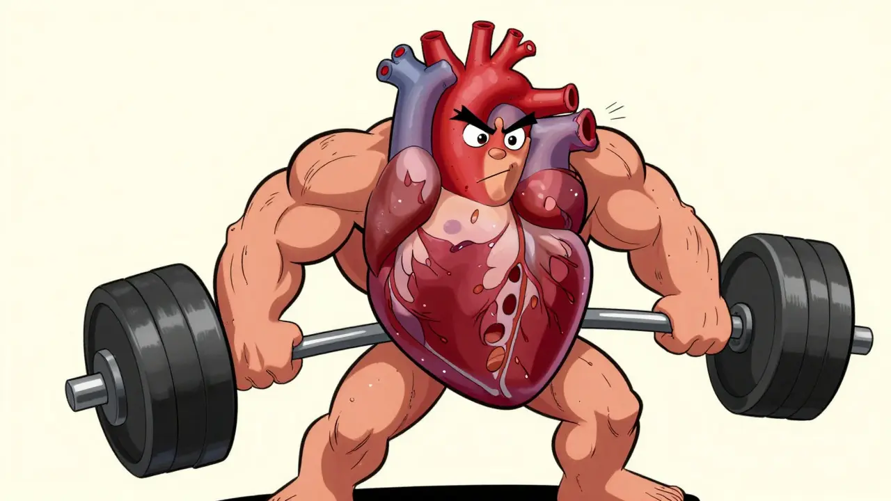
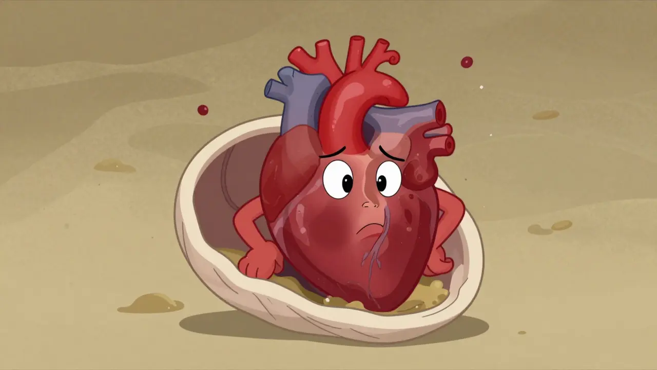
The third major type is the rarest and often the trickiest to diagnose. Restrictive cardiomyopathy (RCM) is A condition where the heart muscle becomes rigid and stiff, impairing filling despite normal pumping strength. Here, the heart doesn’t stretch out, nor does it necessarily get thicker. Instead, it becomes stiff. Think of a water balloon filled with sand-it holds its shape, but it’s hard to squeeze and hard to refill.
In RCM, the heart pumps normally (your ejection fraction might look fine), but it can’t relax enough to fill with blood between beats. This leads to backup pressure in the veins and lungs, causing swelling in the legs and difficulty breathing. The stiffness usually comes from substances infiltrating the heart tissue. Amyloidosis, where abnormal proteins build up, is the most common cause, accounting for about 60% of cases. Other culprits include sarcoidosis, hemochromatosis (iron overload), and Fabry disease.
- Key Sign: Swelling in the abdomen or legs, and persistent fatigue despite resting.
- Diagnostic Challenge: Often mistaken for constrictive pericarditis, requiring cardiac MRI or biopsy for confirmation.
- Treatment Focus: Treating the underlying cause (e.g., removing excess iron or using drugs like tafamidis for amyloidosis).
Because RCM is so rare-only about 1 in 10,000 people-and symptoms overlap with other conditions, diagnosis can take years. Many patients report feeling dismissed because standard tests show a "normal" pumping function. Advanced imaging like cardiac MRI is essential here to spot the subtle signs of infiltration.
How Do Doctors Tell Them Apart?
You might wonder how a doctor knows which type you have. They don’t just guess. They use a combination of tools, primarily echocardiography (an ultrasound of the heart) and cardiac MRI.
| Feature | Dilated (DCM) | Hypertrophic (HCM) | Restrictive (RCM) |
|---|---|---|---|
| Heart Shape | Enlarged, thin-walled | Thickened walls | Normal size, stiff walls |
| Pumping Function | Weak (Low Ejection Fraction) | Normal or Strong | Normal |
| Filling Ability | Impaired due to size | Impaired due to thickness | Severely impaired due to stiffness |
| Primary Cause | Viral, Alcohol, Genetic | Genetic (Sarcomere mutations) | Infiltrative (Amyloid, Iron) |
| Prevalence | Most Common (~50%) | Common (~30-40%) | Rare (~5-10%) |
Genetic testing is also becoming standard, especially for HCM and familial DCM. If you have a relative with cardiomyopathy, getting tested can reveal risks before symptoms ever appear. For example, a 17-gene panel can identify mutations in over 60% of HCM cases. This information helps guide screening for siblings and children.
Living With Cardiomyopathy: What You Can Control
A diagnosis can feel scary, but it’s manageable. The key is sticking to your treatment plan and monitoring your body. For DCM patients, medication adherence is critical. Drugs like SGLT2 inhibitors, once used only for diabetes, now play a huge role in protecting the heart. For HCM patients, staying hydrated and avoiding dehydration during exercise is vital, as low blood volume worsens the obstruction.
Lifestyle changes matter across the board. Limiting salt intake helps reduce fluid buildup, especially in RCM and DCM. Moderate exercise is generally encouraged, but intense competitive athletics may need restriction depending on your risk profile. Always consult your cardiologist before starting a new workout routine.
Don’t ignore symptoms. If you notice increased swelling, sudden weight gain, or worsening shortness of breath, call your doctor. These are signs that your heart is struggling to keep up, and early intervention can prevent hospitalization.
Is cardiomyopathy hereditary?
Yes, many forms are. Hypertrophic cardiomyopathy is strongly genetic, with about 60% of cases caused by inherited mutations. Dilated cardiomyopathy also has a significant genetic component, with 25-35% of cases being familial. Restrictive cardiomyopathy is less commonly inherited directly but can be associated with genetic storage diseases like Fabry disease. If you have a family history, genetic counseling is highly recommended.
Can cardiomyopathy be cured?
There is no universal cure, but many types can be managed effectively. Some causes of restrictive cardiomyopathy, like iron overload, can be treated to halt progression. New targeted therapies for HCM and advanced heart failure drugs for DCM offer significant symptom relief and improved survival. In severe cases, heart transplantation may be considered a curative option.
What are the early warning signs?
Early signs often mimic general fatigue or aging. Look for shortness of breath during mild exertion, swelling in your ankles or feet, rapid heartbeat, dizziness, or chest pain. In children or young adults, fainting during sports is a major red flag for hypertrophic cardiomyopathy. Persistent coughing or difficulty sleeping due to breathlessness are also common indicators.
How does diet affect cardiomyopathy?
Diet plays a supportive role. Reducing sodium intake helps manage fluid retention, which is crucial for all types. Avoiding excessive alcohol is vital, especially for dilated cardiomyopathy, as alcohol can be toxic to heart muscle. A heart-healthy diet rich in vegetables, fruits, and whole grains supports overall cardiovascular health, though it won't reverse structural changes on its own.
When should I see a specialist?
If your primary care doctor suspects heart issues based on symptoms or an abnormal ECG, you should see a cardiologist. Specifically, ask for a cardiomyopathy specialist if you have a family history, unexplained fainting, or if standard treatments aren't working. Specialists have access to advanced imaging like cardiac MRI and genetic testing panels that general cardiologists might not routinely use.